
Figure 2. Effects of trace eyeblink conditioning on survival of newly generated neurons.
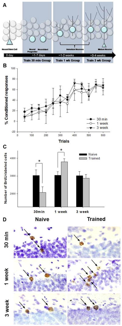
A diagram illustrating the stage of development when BrdU-labeled cells were exposed to training. All animals were injected once with BrdU. They were then trained or not 30min, 1 week or 3 weeks later. To assess the effects of learning on cell survival, animals were euthanized four weeks after the BrdU injection (A). Acquisition of the CR in animals trained 30min, 1 week and 3 weeks after BrdU injection (B). Total number of BrdU-labeled cells in the dentate gyrus in animals trained 30 min, 1 week or 3 weeks after injection compared to naïve animals. Trace conditioning increases the number of BrdU-labeled cells when rats are trained 1 week after injection, but decreases the number that survive when they were trained just 30 min after injection (C). Photomicrographs at 100x of BrdU-positive cells (arrows) from each experimental condition. Cells are located along the infrapyramidal blade in the dorsal hippocampus. Images represent the relative differences in the number of BrdU-labeled cells in response to learning when compared to naïve animals. The granularity in some cells depicts the extended time interval between the BrdU injection and sacrifice (D). * indicates p < 0.05.