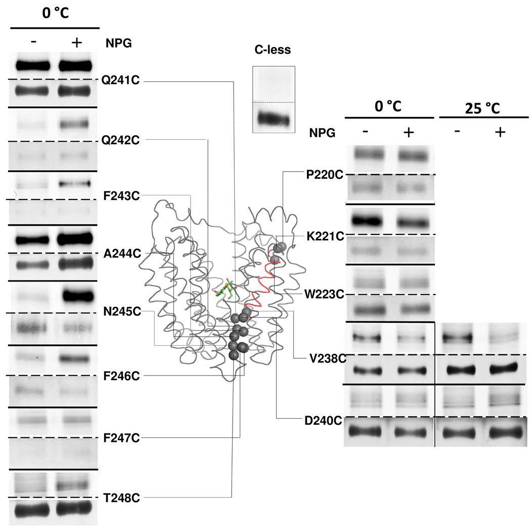
Figure 1. Labeling of single-Cys replacements in helix VII.
RSO vesicles containing given single-Cys LacY mutants (0.1 mg of total protein) were incubated with 40 µM TMRM for 30 min at 0 °C or 25 °C in the absence or presence of 1 mM αNPG as indicated. Reactions were terminated by addition of 10 mM DTT. The membranes were solubilized with 2% DDM and biotinylated LacY was purified with monomeric avidin, and then subjected to SDS-PAGE. LacY bands labeled with TMRM (upper panels) and silver-stained (lower panels) were imaged. The Cα atoms of labeled single-Cys mutants are shown as gray spheres superimposed on the backbone of LacY [PDB ID: 1PV7]. LacY is viewed perpendicular to the membrane with the N-terminal helix bundle on the left and the C-terminal helix bundle on the right. αNPG is shown as a stick model at the apex of the inward-facing cavity. Helix VII is highlighted in red. TMRM labeling of C-less LacY for 30 min at 0°C is shown as the negative control.

