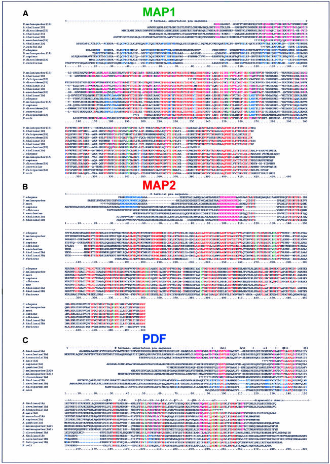
Fig. 1. Alignment of the amino acid sequences of PDF and MAP from higher eukaryotes. The sequences of MAP and PDF (see text) were used as input probes with the help of the BLAST software (Altschul et al., 1990) to search data at the web site of the Arabidopsis Information Resource (http://www.arabidopsis.org/cgi-bin/blast/TAIRblast.pl) and at the NCBI. Matching EST sequences were compared with each other and assembled when overlapping. The genomic fragments were analysed with the various software predicting intron–exon splice sites (Hebsgaard et al., 1996) and, by using the amino acid sequence homologies, assembled and compared with the ESTs. Arabidopsis thaliana cDNAs were cloned and sequenced. The deduced amino acid sequence is shown. Translated sequences were aligned with Clustalx (Jeanmougin et al., 1998) and then manually. A series of question marks indicates that the corresponding nucleotide sequence is partial or missing. The numbering of each of the three amino acid sequence sets is shown below each block of 150 residues. Amino acids in italic at the N-terminus indicate putative alternative translation starts. Residues strictly conserved within the catalytic core are shown in red or in green for the ligands of the catalytic metal. (A) MAP1 alignments. The amino acid similarity between groups is shown in blue for cytoplasmic MAP (bottom) or in pink for organellar MAP (top). (B) MAP2 alignments. The deduced amino acid sequence at the 3′ end of the EST of the Bombyx mori sequence was modified because of an obvious frame-shift error. The poly-basic common amino acid stretch is shown in pink. The second poly-basic sequence found in animal sequences is in blue. (C) PDF alignments. The additional similarity shared by plant PDF1A (top) is coloured in pink. Specific features of PDF1B types (bottom) are shown in blue. Secondary structure elements are indicated on top.

An official website of the United States government
Here's how you know
Official websites use .gov
A
.gov website belongs to an official
government organization in the United States.
Secure .gov websites use HTTPS
A lock (
) or https:// means you've safely
connected to the .gov website. Share sensitive
information only on official, secure websites.
