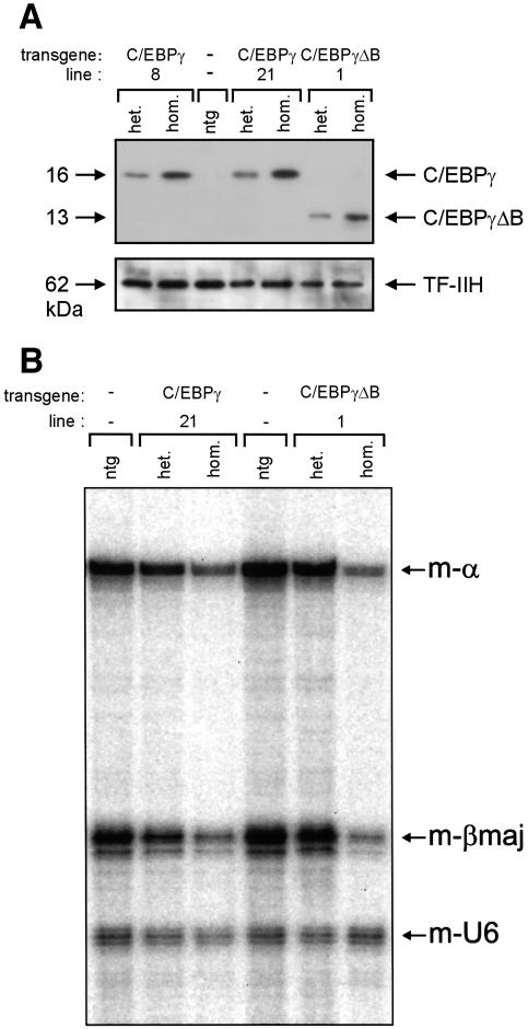
Fig. 4. High levels of the EBP proteins result in reduced expression of the α- and β-globin genes. (A) Ten micrograms of E12.5 fetal liver nuclear extract were used for western blot analysis of EBP protein expression. Results are shown for non-transgenic (ntg), heterozygous (het.) and homozygous (hom.) EBP transgenics of the lines indicated. The polyclonal rabbit antiserum used was raised against the N-terminal part of C/EBPγ that is shared by the C/EBPγ and C/EBPγΔβ proteins. Note that the levels of endogenous C/EBPγ, although present in the RT–PCR analysis (Figure 1D), are too low for detection with this antiserum. A mouse monoclonal antibody recognizing the p62 subunit of TF-IIH was used as a loading control. (B) E12.5 livers were dissected from non-transgenic (ntg), heterozygous (het.) and homozygous (hom.) EBP transgenics of the lines indicated and used for RNA isolation. Approximately 1 µg of total RNA was used for quantitative S1 nuclease analysis of mouse α- (m-α) and βmajor-globin gene expression (m-βmaj); a probe detecting mouse U6 RNA (m-U6) was used as an internal loading control (Wijgerde et al., 1996).

An official website of the United States government
Here's how you know
Official websites use .gov
A
.gov website belongs to an official
government organization in the United States.
Secure .gov websites use HTTPS
A lock (
) or https:// means you've safely
connected to the .gov website. Share sensitive
information only on official, secure websites.
