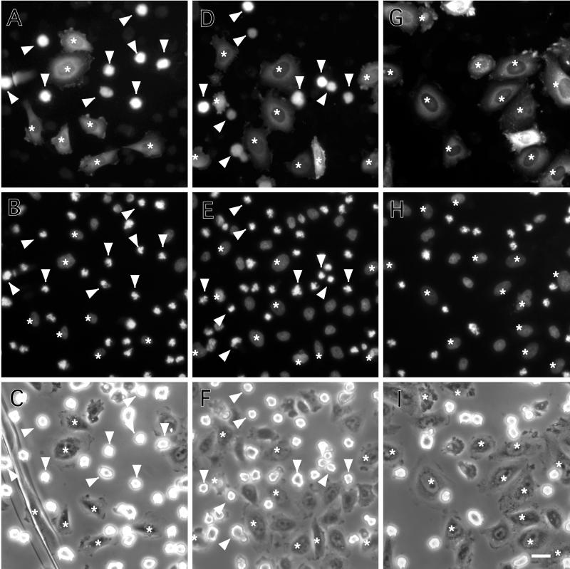
Figure 3.
Delay of M-phase entry by microinjection of 7th mutant. CHO cells were microinjected with buffer alone (A–C), wild-type caldesmon (D–F), or 7th mutant caldesmon (G–I). FITC-dextran was coinjected to identify injected cells (A, D, and G). Cells were then synchronized for cell division by thymidine treatment followed by nocodazole treatment as described in MATERIALS AND METHODS. After 4 h of nocodazole treatment, cells were fixed and stained with DAPI to examine chromosome condensation (B, E, and H). (C, F, and I) Corresponding phase-contrast images of the same fields. Arrowheads and asterisks indicate mitotic and nonmitotic cells, respectively. Note that none of cells injected with 7th mutant caldesmon were in mitosis as judged by their flat morphology (G), and by DAPI staining (H). On the contrary, about half of cells injected with either buffer alone (A–C) or wild-type caldesmon (D–F) went into mitosis as judged by their morphology (A and D) and by chromosome condensation (B and E). Bar, 25 μm.

