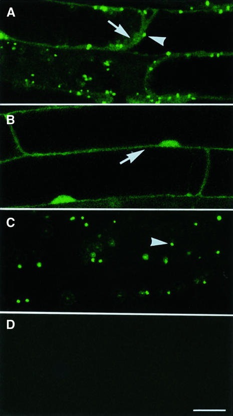
Fig. 5. Subcellular localization of GFP–PTS1 fusion protein in ped2 mutant. Seedlings were grown under continuous illumination for 10 days. Images of the green fluorescence derived from GFP in root cells were taken by a confocal laser microscope as single optical sections. (A) Subcellular localization of GFP–PTS1 expressed in the cells of a ped2 mutant. (B) Subcellular localization of GFP expressed in the cells of a ped2 mutant. (C) Subcellular localization of GFP–PTS1 expressed in the cells of a wild-type plant. (D) No fluorescence was observed in non-transformed cell of a ped2 mutant. Arrowheads indicate fluorescence detected in peroxisomes, whereas the arrows indicate fluorescence detected in the cytosol. Bar in (D), 20 µm. Magnifications of (A)–(D) are the same.

An official website of the United States government
Here's how you know
Official websites use .gov
A
.gov website belongs to an official
government organization in the United States.
Secure .gov websites use HTTPS
A lock (
) or https:// means you've safely
connected to the .gov website. Share sensitive
information only on official, secure websites.
