
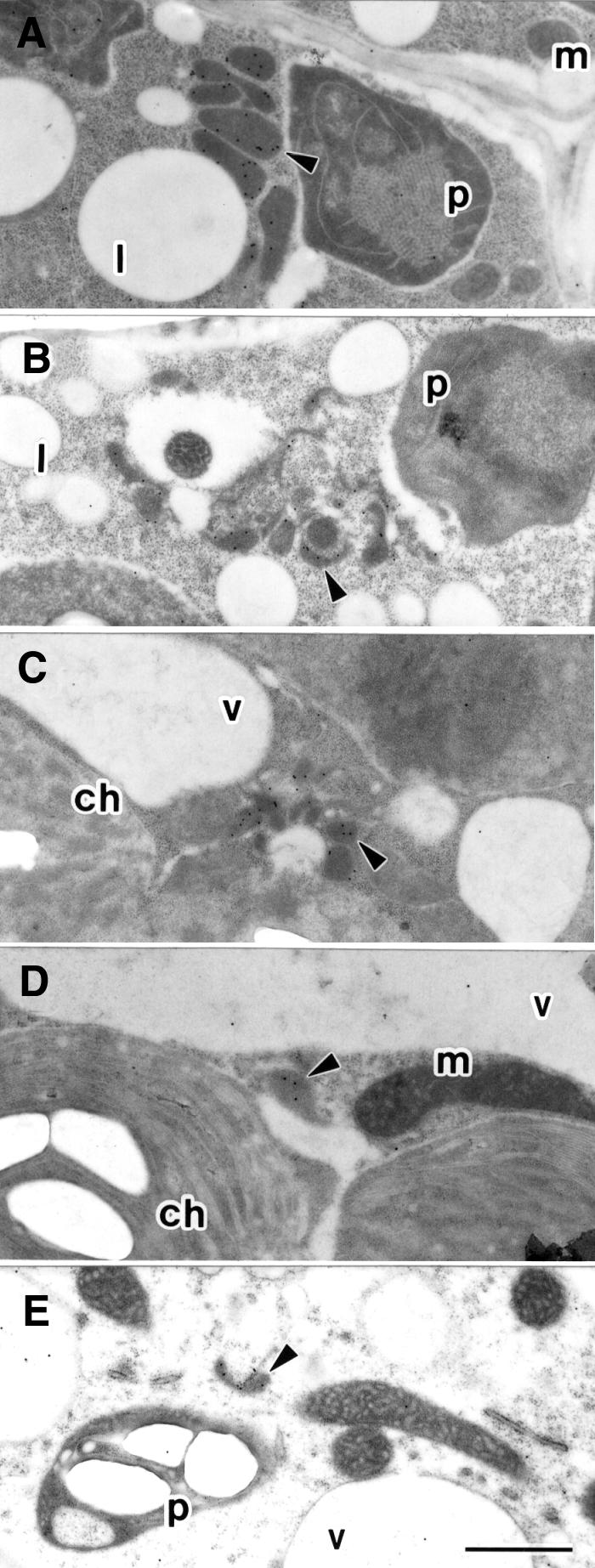
Fig. 7. Electron microscopic analysis of peroxisomes in the cells of the ped2 mutant. (A) Etiolated cotyledon of wild-type Arabidopsis, grown for 5 days in darkness, was stained with 3-ketoacyl CoA thiolase-specific antibody. (B) Etiolated cotyledon of ped2 mutant, grown for 5 days in darkness, was stained with 3-ketoacyl CoA thiolase-specific antibody. (C) Green cotyledon of ped2 mutant, grown for 7 days under continuous illumination, was stained with hydroxypyruvate reductase-specific antibody. (D) Leaf of ped2 mutant, grown for 14 days under continuous illumination, was stained with hydroxypyruvate reductase-specific antibody. (E) Root of ped2 mutant, grown for 14 days under continuous illumination, was stained with catalase-specific antibody. Arrowhead, peroxisome; m, mitochondrion; l, lipid body; p, plastid; ch, chloroplast; v, vacuole. Bar in (E), 1 µm. Magnification of (A)–(E) is the same.