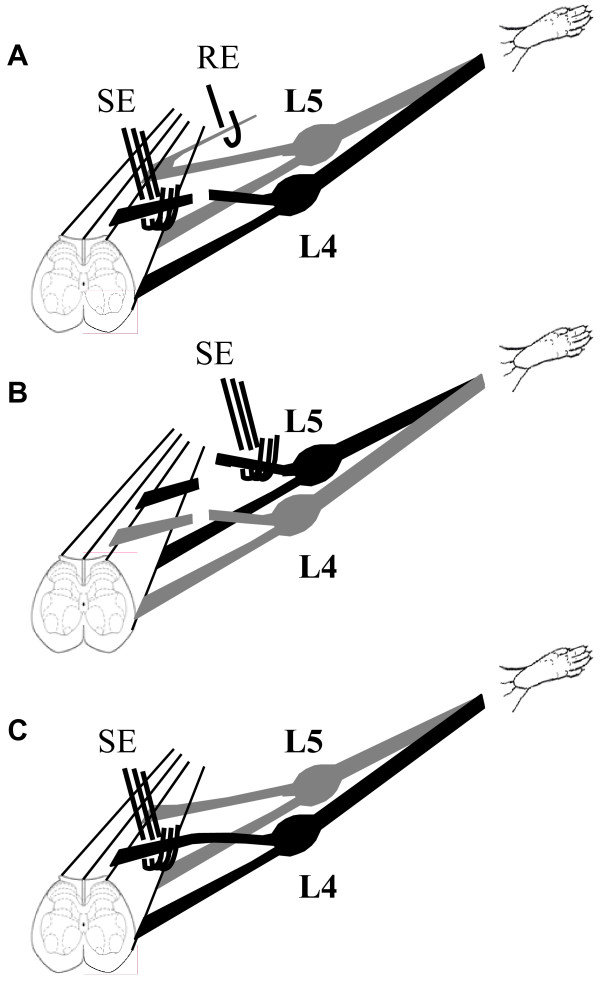
Figure 1.
Diagram of the experimental setup. A. The left L4 dorsal root is cut. The central stump is placed over a stimulating electrode (SE) while placing a strand from the left L5 dorsal root on a recording electrode (RE). B. A second cut is made at the left L5 dorsal root. The peripheral stump of the left L5 dorsal root is placed over a stimulating electrode. Seven minutes after Evans Blue injection (i.v.), electrical stimulation (20 V, 5 Hz, 0.5 ms for 5 min) is delivered, while a series of images is taken. C. A setup for stimulating an intact dorsal root.

