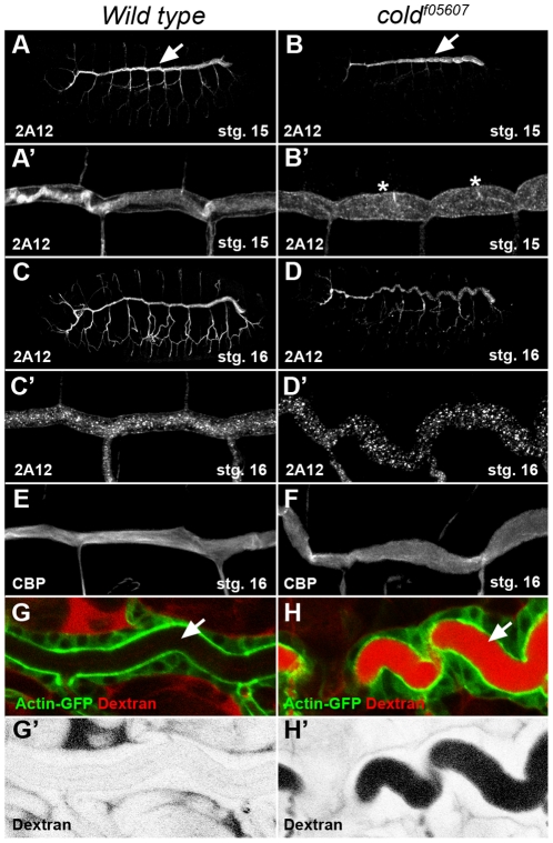
Figure 2. The tracheal morphology and paracellular barrier integrity are perturbed in cold embryos.
(A–D) Projections of confocal stacks corresponding to wild type and coldf05607 embryos staged as indicated and stained for the 2A12 tracheal luminal antigen. The same trachea are shown at higher magnification in panels A′–D′. At stage 15, the morphology of the tracheal dorsal trunk (arrows) is affected and displays a series of cysts (asterisks) visible in cold mutants. By stage 16, the dorsal trunk adopts a convoluted shape. (E–F) Projections of confocal stacks showing the tracheal dorsal trunk stained with fluorescent chitin binding probe (CBP). In the wild type, the chitin cable displayed a fibrous structure that was lost in cold embryos of the same stage. (G,H) Single confocal sections showing a view of the dorsal tracheal trunk marked by ActinGFP (green) and corresponding to stage 16 live embryos injected in the hemolymph with rhodamine 10 kDa dextran (red). (G′,H′) show a greyscale negative image of the red channel shown in G,H.

