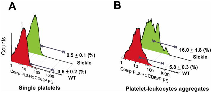
Figure 2. Increased P-selectin on platelet-leukocyte aggregates in sickle mice.
Platelets and leukocytes were recognized by their forward and side scatter characteristics. Platelet singlets were further identified with anti-GPIX-FITC. Platelet-leukocyte complexes were identified by anti-CD45-PerCP-Cy5.5 and anti-GPIX-FITC positive fluorescence. P-selectin expression was assessed using anti-CD62P-PE. Representative histograms showing P-selectin expression on (A) platelet singlets and (B) platelet-leukocyte heteroaggregates, n ≥ 5. Data are expressed as means ± SD.

