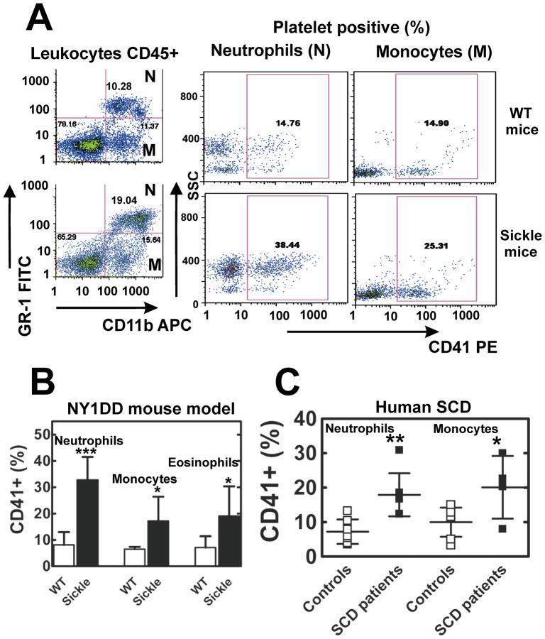
Figure 3. Increased platelet interactions with leukocytes in NY1DD sickle mice and SCD patients.
(A) Representative FACS analysis of whole blood platelet-neutrophil (N) and platelet-monocyte (M) aggregates in WT and SCD mice. CD45+ leukocytes: neutrophils (N) and monocytes (M) were gated by forward and side light scatter (not shown) and the differential expression of GR-1-FITC, and CD11b-APC. The percentage of neutrophils and monocytes that are found in heteroaggregates with platelets (CD41+) is greater in SCD than WT mice. (B) Quantitative analysis of CD41+ neutrophils, monocytes and eosinophils in WT and sickle mice blood. Results are presented as means ± SD, n=10 for both groups, ***p<0.001, *p<0.05. (C) Increased formation of platelet-neutrophil and platelet-monocyte aggregates in blood obtained from the adult SCD patients compared to healthy controls. Results are presented as means ± SD, n=7 for both groups, ** p<0.005, *p<0.05.

