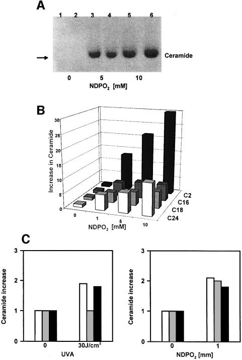
Fig. 4. Singlet oxygen generates ceramide in sphingomyelin-containing liposomes. (A) HPTLC of NDPO2-induced ceramide formation in liposomes. Liposomes were treated with 0 (lanes 1 and 2), 5 (lanes 3 and 4) and 10 mM NDPO2 (lanes 5 and 6), and chloroform/methanol lipid extracts prepared immediately thereafter and analysed. Iodine vapour was used to stain ceramides. (B) Analysis of NDPO2-induced ceramide formation in liposomes by HPLC/mass spectroscopy. Liposomes were treated for 30 min with increasing doses (0–10 mM) of NDPO2 and lipid extracts prepared immediately thereafter. (C) Analysis of ceramide in long-term cultured human keratinocytes by HPLC/mass spectroscopy. Lipid extracts were prepared 30 min after stimulation of cells with 30 J/cm2 UVA or 1 mM NDPO2. C24, white bars; C18, grey bars; C16, black bars.

An official website of the United States government
Here's how you know
Official websites use .gov
A
.gov website belongs to an official
government organization in the United States.
Secure .gov websites use HTTPS
A lock (
) or https:// means you've safely
connected to the .gov website. Share sensitive
information only on official, secure websites.
