
Figure 3.
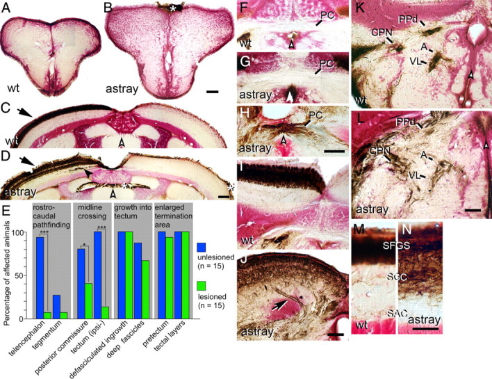
Correction and recurrence of errors by regenerating optic axons in adult astray mutants. Optic axons are stained brown in cross sections of the adult brain (counterstained in red); dorsal is up. White arrowheads indicate the brain midline. Asterisks indicate nonspecific labeling of the meninges. A, B, No regenerated optic axons are present in wild-type (A) or astray (B) telencephalon. C, D, The regenerated optic projection in the tectum is exclusively contralateral (arrow) in wild type (C) and astray (D), but erroneous growth of deep fascicles (black arrowhead in D) recurs in astray. E, Frequencies of different astray phenotypes before and after regeneration of the optic projection in animals preselected for the presence of telencephalic tracts in larvae. *p < 0.05, ***p < 0.0001. F–H, Regenerating optic axons do not cross the posterior commissure (PC) in wild-type fish (F). Regenerated optic axons show ectopic crossing in the posterior commissure in some astray animals (H), but not in others (G). I, J, Regenerated axons enter the tectum in separate fascicles in astray (arrow in J), but not in wild type (I). K, L, Termination areas of regenerated optic axons in the pretectum are expanded in astray animals (L), compared with wild-type animals (K). M, N, In astray (N), reinnervation of tectal layers is expanded compared with wild type (M) after regeneration. For abbreviations see Figure 2. Scale bars: A, B, 200 μm; C, D, F–L, 100 μm; M, N, 50 μm.