
Figure 4.
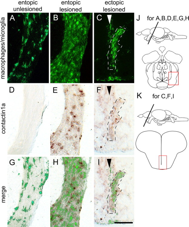
Deafferented ectopic optic tracts in the telencephalon of astray mutants display macrophage/microglial cell activation and increased contactin1a mRNA expression comparable to entopic tracts. Cross sections through the adult brain are shown as indicated in J and K. Macrophage/microglial cell immunolabeling (A–C) and contactin1a mRNA labeling (D–F) is comparable between deafferented entopic (B, E, H) and ectopic astray optic tracts (C, F, I). Both signals are increased compared with unlesioned entopic tracts (A, D, G). Arrowheads in C, F, and I indicate telencephalic midline. G–I show superimposition of macrophage/microglial cell and contactin1a mRNA labeling. Scale bar, 200 μm.