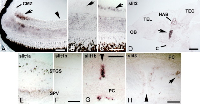
Figure 5.
Robo2 and slits are expressed during regeneration of the adult optic projection. Cross sections are shown, except for D. A, In the retina of unlesioned juvenile, 4-week-old animals, robo2 mRNA is expressed in recently differentiated retinal ganglion cells in the peripheral growth zone of the retina (arrow) next to the ciliary margin zone (CMZ). Older, more central retinal ganglion cells (arrowhead) do not express detectable levels of robo2 mRNA. B, C, In the adult (>3 months of age) central retina, robo2 mRNA is reexpressed in the retinal ganglion cell layer at 2 weeks postlesion (arrow in C) compared with the retinal ganglion cell layer in unlesioned controls (arrow in B). D, A sagittal section of the brain is shown (rostral left, dorsal up). Conspicuous expression of slit2 mRNA is found in the habenula (HAB) and in the ventral diencephalon (arrow) at the level of the optic chiasm (C). OB, Olfactory bulb; TEL, telencephalon; TEC, tectum mesencephali. E, F, Slit1a (E), but not slit1b (F), is expressed in the deafferented tectum at 1 week postlesion. SPV, stratum periventriculare; SFGS, stratum fibrosum et griseum superficiale. G, Strong local expression of slit1b mRNA is found at the level of the posterior commissure (PC) in cross sections of the brain. H, Low levels of slit3 mRNA expression are found in the pretectum, including the PPd area (arrow). Arrowheads in G and H indicate the brain midline. Scale bars: A, 50 μm; B, C, 50 μm; D, 200 μm; E, F, 100 μm; G, 50 μm; H, 100 μm.

