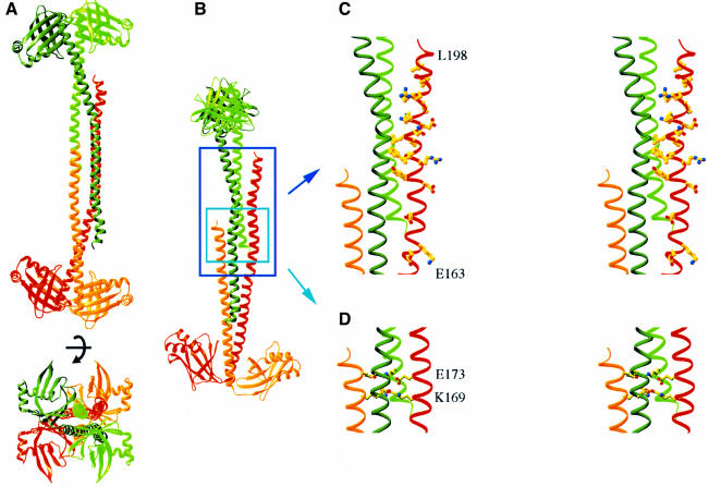
Fig. 3. Structure of the Xrcc4 tetramer. (A) Two orthogonal views of an Xrcc4 tetramer. The crystallographic dyad axis is marked. The two dimers are colored in green and red; a darker color for the L subunit and a lighter color for the S subunit. (B) A view that shows the interdigitation of the two Xrcc4 dimers. (C) Stereo drawings of the conserved residues between 170 and 203 in one L subunit of the Xrcc4 tetramer and (D) two layers of repeated salt bridges in the four-helix bundle region. Both are viewed in the same orientation as in (B).

An official website of the United States government
Here's how you know
Official websites use .gov
A
.gov website belongs to an official
government organization in the United States.
Secure .gov websites use HTTPS
A lock (
) or https:// means you've safely
connected to the .gov website. Share sensitive
information only on official, secure websites.
