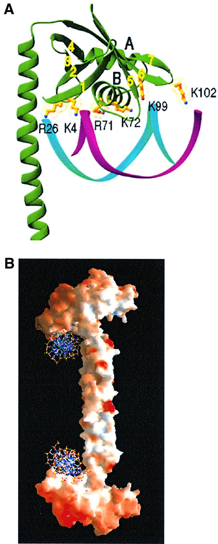
Fig. 5. A model of DNA binding by Xrcc4. (A) Basic residues of Xrcc4, which are potentially involved in DNA binding, are shown together with a B-DNA. The αB helix of the HTH motif faces the major groove and interacts with the DNA backbone. (B) A molecular surface presentation of Xrcc4 shown with positive (blue) and negative (red) electrostatic potential. Two DNA molecules are modeled to bind the S subunits as shown in (A) at the base of the head domain, as one of many possible ways in which the protein and DNA can associate with one another.
