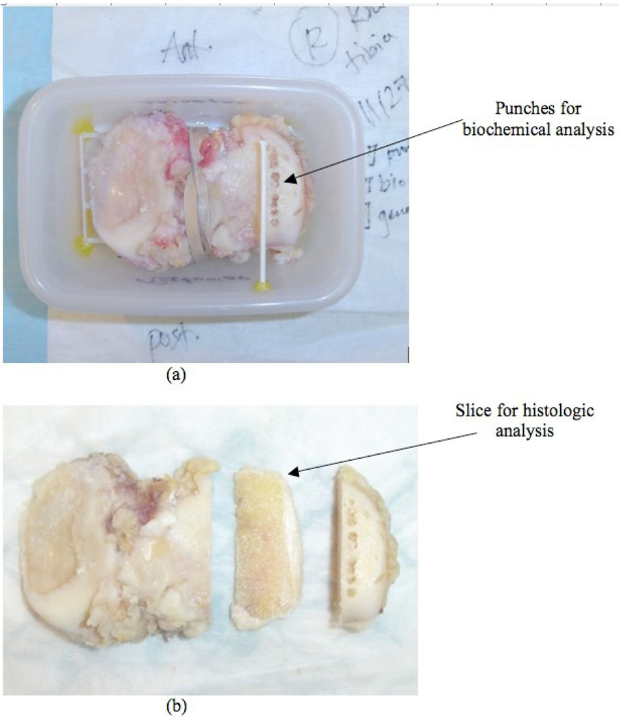
Figure 2.
Specimen preparation. The specimen was mounted on a plastic grid for location reference, and then placed and glued into a plastic container and immersed in phosphate-buffered saline (PBS) for ex vivo MRI (a). An additional thin plastic tube marker was placed on top of the location where the histology slice was obtained after the MRI. After MRI, punches were taken for biochemical analysis (a) and a 3mm histology slice was cut next to the biochemical punches for histological analysis (b).

