
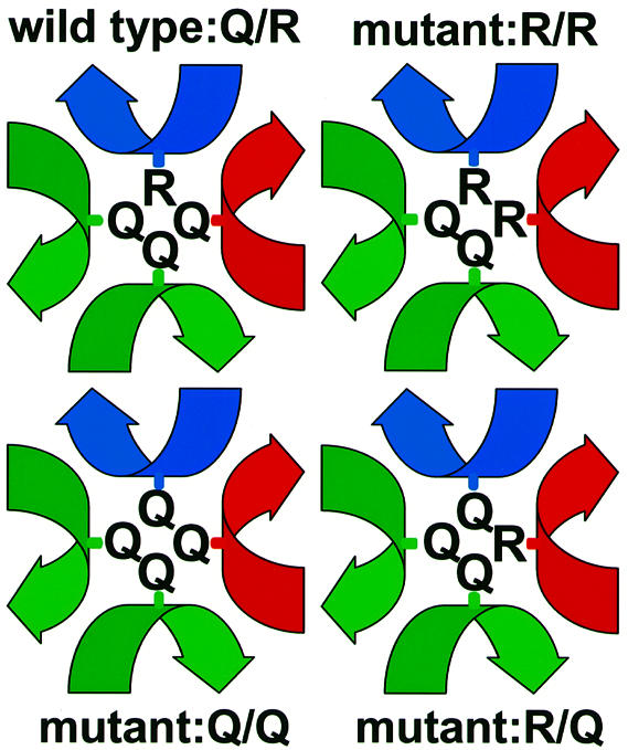
Fig. 1. Schematic illustration of the arrangement of amino acid side chains involved in the ‘0’ layers of wild-type and mutant SNARE complexes used in this study. Local helical axes of the SNARE proteins are displayed in blue for Snc2p, red for Sso2p and green for Sec9p.