Fig. 3.

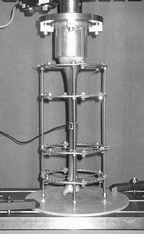
Experimental set-up for analysis of the compressive loads in the osteotomy at indirect weight loading; the connecting rods rest on the base plate, leaving the distal bone end levitating; the loading cell covers the defect completely

Experimental set-up for analysis of the compressive loads in the osteotomy at indirect weight loading; the connecting rods rest on the base plate, leaving the distal bone end levitating; the loading cell covers the defect completely