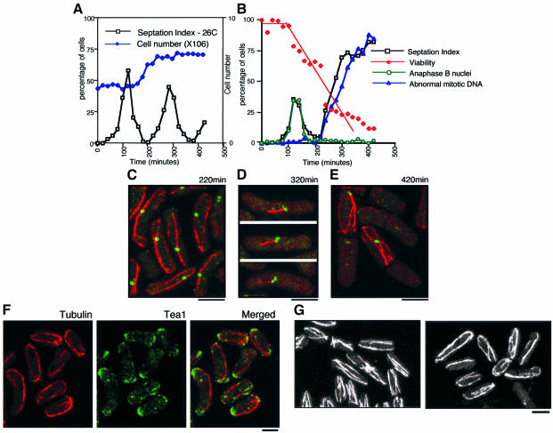
Fig. 3. Alp4 is required for both the formation of mitotic bipolar spindles and the integrity of interphase cytoplasmic microtubules. (A and B) Centrifugal elutriation. Small early G2 cells of an alp4-1891 strain (DH1891) were collected by centrifugal elutriation and the cultures were divided into two parts, one part incubated at 26°C (A) and the other at 36°C (B). Samples were collected at 20 min intervals, and cell number (open diamonds in dark blue), septation index (open squares in black) and viability (open diamonds in red, the percentage of viable colonies) were measured. Immunofluorescence microscopy with anti-tubulin, anti-Sad1 antibody and DAPI was performed to examine the percentage of cells containing anaphase B nuclei (open circles in green) and abnormal mitotic DNA (open triangles in blue). (C) Abnormally long interphase microtubules in the alp4-1891 mutant. Confocal microscopy was performed using synchronous interphase samples (220 min at 36°C) with anti-tubulin (red) and anti-Sad1 (green) antibodies. Merged images are shown. (D) Aberrant mitotic spindles. Characteristic cells showing monopolar-like spindles (320 min) are presented. (E) Displaced long interphase microtubules. Interphase-like cells with longer cytoplasmic microtubules after an aberrant mitosis (420 min) are shown. (F) Tea1 localization in the alp4-1891 mutant. alp4 mutants were incubated at 26°C for 3 h in the presence of 10 mM HU and shifted up to 36°C after washout of HU. After 2.5 h incubation, cells were fixed and processed for immunofluorescence microscopy using anti-tubulin (left) and anti-Tea1 (middle) antibodies. Merged images are shown on the right. (G) Defects in microtubules by overexpression of alp4+. Cells containing integrated nmt1-alp4+ were grown in the absence of thiamine, and processed for immunofluorescence microscopy using anti-tubulin antibody. Images are from confocal microscopy. Cells from a wild-type control (left) and alp4+ overexpression after 16 h (right) are shown. The bar indicates 10 µm.

An official website of the United States government
Here's how you know
Official websites use .gov
A
.gov website belongs to an official
government organization in the United States.
Secure .gov websites use HTTPS
A lock (
) or https:// means you've safely
connected to the .gov website. Share sensitive
information only on official, secure websites.
