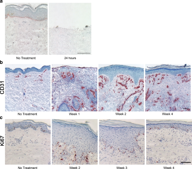
Fig. 3.
Vascular and epidermal changes induced by CO2 laser wounding in human forearm skin. a CO2 laser creates a partial thickness wound by vaporizing the entire epidermis and the upper part of the papillary dermis. Basement membrane is highlighted by lamin-γ2 staining for reference. Scale bar, 100 μm. b Time course of blood vessel formation after wounding in human skin. Endothelial cells are stained with CD31. Blood vessel formation starts within the first week post-wounding, and hyper-vascularization is visible for at least 4 weeks. Scale bar, 100 μm. c Time course of re-epithelialization of partial thickness wounds in human skin. Proliferative cells are stained with Ki67. Epidermal hyperplasia is visible at weeks 2 and 3, and epidermal thickness is normalized by week 4 (also visible on b). Scale bar, 100 μm

