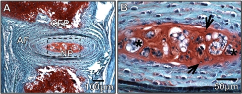
Fig. 1.
Morphology and Composition of the Intervertebral disc. a Histological sections of the IVD from a 4 week old mouse stained with safranin-O/fast green demonstrates IVD morphology and regionalization of the nucleus pulposus (NP), annulus fibrosis (AF), and cartilaginous end-plate (CEP). Proteoglycan content is indicated by red stain (scale bar represents 100 uM). b Enlarged view of the nucleus pulposus region in A. Arrows indicate large aggregations of vacuolated notochord cells and * indicate the smaller more disperse cartilage-like cells (scale bar represents 50 uM)

