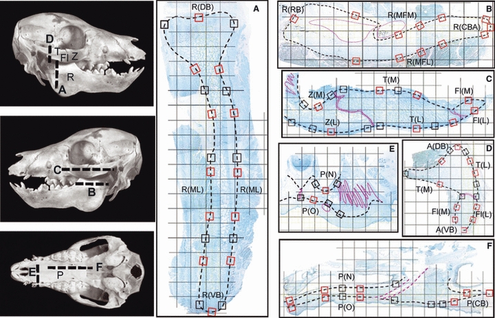
Fig. 1.

Left, photographs of piglet skulls indicating planes of section for (A–F). The right mandibular ramus and zygomatic arch were sectioned coronally, and the left were sectioned horizontally. Fl, flange of the zygomatic bone; P, palate; R, mandibular ramus; T, temporal bone; Z, zygomatic bone. (A–F) Examples of sections (composite images) overlaid by a 2.3-mm grid. The small boxes are the 89 1-mm locations examined for periosteal appositional or resorptive activity; those outlined in red were also analyzed quantitatively and grouped into regions (see Table 1 for list of region abbreviations). Dotted pink lines indicate sutures and neurovascular bundles. (A) Coronal section through the condylar region of the ramus showing dorsal and ventral borders, R(DB) and R(VB), and ramal surfaces, R(ML). (B) Horizontal section through the ramus showing rostral and caudal borders, R(RB) and R(CBA), and lateral and medial surfaces of the mandibular foramen area, R(MFL) and R(MFM). (C) Horizontal section through the arch showing medial (M) and lateral (L) surfaces of the zygomatic (Z), zygomatic flange (Fl) and temporal (T) bones. (D) Coronal section through the articular region of the arch showing dorsal and ventral borders of the arch, A(DB) and A(VB), as well as lateral and medial surfaces of the temporal bone and zygomatic flange. (E) Coronal section through the hard palate showing nasal and oral surfaces, P(N) and P(O). (F) Parasagittal section through the palate showing the caudal border, P(CB), as well as nasal and oral surfaces.
