
Fig. 3.
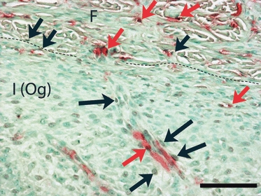
Coronal section from the lateral surface of the temporal bone, double-stained for lectin (Vector Red) and BrdU (DAB/Ni, brown). A dashed line separates the fibrous (F) and inner osteogenic [I (Og)] layers of periosteum. This region is strongly appositional. Double-labeled cells (red arrows) are seen in both layers of periosteum, indicating replication of endothelial cells, probably associated with angiogenesis. In addition, many cells labeled only with BrdU (black arrows) are located one or two cell layers distant from lectin-labeled vessels. These may be pericytes. Calibration bar: 100 μm.