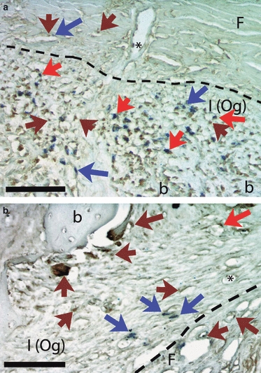
Fig. 5.

Coronal section through the temporal bone, double-labeled for CTR (calcitonin receptor, Nova Red, which appears as dark red/brown) and BrdU (NBT/BCIP, blue). Dashed lines separate the fibrous (F) and inner [I (Og)] layers of periosteum. (a) Appositional lateral surface. CTR label (brown arrows) is plentiful in the inner layer of periosteum, as is BrdU label (blue arrows), and many cells are double-labeled (red arrows). In the fibrous layer CTR label is seen in cells associated with the vasculature, including one attached to the inner wall of a vessel (asterisk). (b) Resorptive medial surface. CTR label (brown arrows) is prominent in active osteoclasts at the bone surface and in mononucleated cells nearby. BrdU-labeled cells (blue arrows) are less numerous than in (a) and are associated with the vasculature, including one that is double-labeled (red arrow) and one apparently inside a vessel (asterisk). Calibration bars: 100 μm. b, bone.
