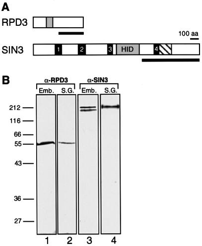
Fig. 1. SIN3 and RPD3 polyclonal antibodies are highly specific. (A) Schematic diagrams of SIN3 and RPD3 proteins. Solid bars indicate regions used as antigens for generating polyclonal antibodies. The RPD3 region does not include the deacetylase domain, indicated by a shaded box. The SIN3 region contains paired-amphipathic helix (PAH) 4, indicated by a solid box, and a conserved region of undefined function, indicated by a hatched box, but does not include PAH1–3 or the histone deacetylase interaction domain (HID). (B) Western blots of total protein extracted from 0–12 h Drosophila embryos (Emb.) and from larval salivary glands (S.G.) were probed with purified RPD3 antibody (lanes 1 and 2) or SIN3 antibody (lanes 3 and 4). The positions of protein molecular weight size markers are indicated on the left.

An official website of the United States government
Here's how you know
Official websites use .gov
A
.gov website belongs to an official
government organization in the United States.
Secure .gov websites use HTTPS
A lock (
) or https:// means you've safely
connected to the .gov website. Share sensitive
information only on official, secure websites.
