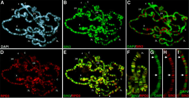
Fig. 2. SIN3 and RPD3 co-localize throughout euchromatin but are absent from heterochromatin. (A–E) A single polytene chromosome spread stained for both SIN3 and RPD3 and counterstained with DAPI. In (E), the diamond and sphere indicate loci that stain predominantly for SIN3 and RPD3, respectively. (F) Higher magnification image of a spread co-stained for SIN3 and RPD3. (G–I) Higher magnification images of another spread stained for SIN3 and counterstained with DAPI. Arrows highlight the non-overlapping pattern of SIN3 and DAPI. Co-localization of two antibodies appears as yellow fluorescence. Chromosome arms (X, 2L, 2R, 3L, 3R and 4) are indicated at the tip, and the chromocenter is indicated by ‘C’. In (A–E), the chromocenter is broken into two pieces. Antibodies used for staining are indicated at the bottom of each panel. The color of the lettering matches the color of the fluorescence.

An official website of the United States government
Here's how you know
Official websites use .gov
A
.gov website belongs to an official
government organization in the United States.
Secure .gov websites use HTTPS
A lock (
) or https:// means you've safely
connected to the .gov website. Share sensitive
information only on official, secure websites.
