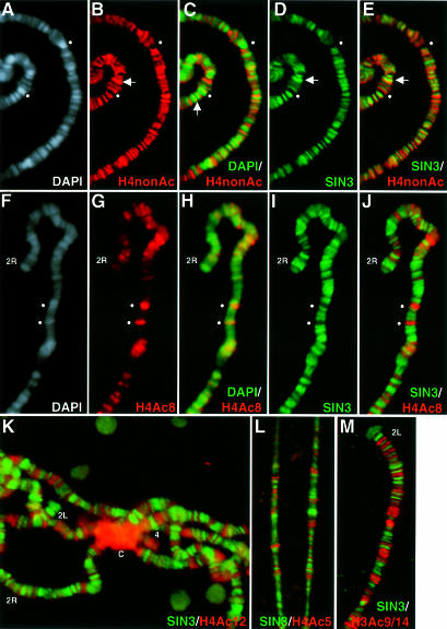
Fig. 3. Chromosomal sites of SIN3 binding and histone hyperacetylation are mutually exclusive. (A–E) A section of a single polytene chromosome spread stained for both H4nonAc and SIN3. Horizontal arrows indicate a region with strong H4nonAc and SIN3 binding. The vertical arrow indicates binding of H4nonAc at the band–interband junction. (F–J) A section of a single polytene chromosome spread stained for both SIN3 and H4Ac8. In (A–J), spheres indicate loci that stain strongly with DAPI and α-H4Ac8, but do not stain with α-H4nonAc or α-SIN3. (K–M) Polytene chromosome spreads co-stained for SIN3 and H4Ac12, H4Ac5 or H3Ac9/14, respectively. Co-localization of two antibodies appears as yellow fluorescence. Chromosome arms (2L, 2R and 4) are indicated at the tip and the chromocenter is indicated by ‘C’ in (K). Antibodies used for staining are indicated at the bottom of each panel. The color of the lettering matches the color of the fluorescence.

An official website of the United States government
Here's how you know
Official websites use .gov
A
.gov website belongs to an official
government organization in the United States.
Secure .gov websites use HTTPS
A lock (
) or https:// means you've safely
connected to the .gov website. Share sensitive
information only on official, secure websites.
