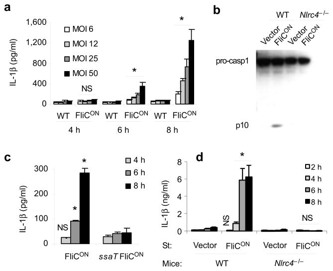
Figure 1. Characterization of flagellin-expressing S. typhimurium.
(a–d) BMDM were infected with ST-WT or ST-FliCON under conditions where SPI2 T3SS is expressed and SPI1 T3SS is not expressed at the indicated multiplicity of infection (MOI) for one hour followed by treatment with gentamicin. Total infection time is indicated. (a) IL-1β concentration determined by ELISA. (b) Caspase-1 processing analyzed by immunobloting. (c) IL-1β concentration after infection with ST-FliCON or SPI2 mutant ST-FliCON (ssaT) at MOI 12. (d) IL-1β concentration after infection of WT or Nlrc4−/−BMDM at MOI 10. Data is representative of at least three experiments. * = p < 0.05, NS = p > 0.05 from relevant control.

