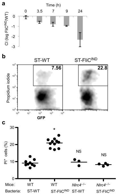
Figure 4. Evidence for pyroptosis in vivo.
(a–c) Mice were infected with either ST-WT (flgB GFP) or flagellin-inducible ST-FliCIND (flgB GFP pEM87) for 48 h, after which doxycycline was injected to induce flagellin expression. (a) Competitive index of ST-FliCIND/ST-WT after synchronized induction of flagellin expression (n=2, except 0h n=3). (b–c) GFP-containing cells from the peritoneal wash were analyzed by flow cytometry for membrane integrity by propidium iodide staining (PI). (b) Representative flow cytometry analysis and (c) percent PI positive bacteria containing cells from WT (n=9 and n=11 for ST-WT and ST-FliCIND) or Nlrc4−/− (n=3 and n=4 for ST-WT and ST-FliCIND) mice per group, average is shown (bar). * = p < 0.001, NS = p > 0.05.

