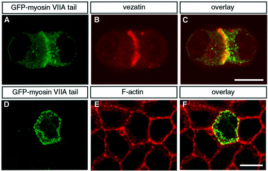
Fig. 3. Co-localization of endogenous vezatin and the myosin VIIA tail fused to GFP in transfected MDCK cells. As soon as cell contacts can be detected, the myosin VIIA tail (A) co-localizes with endogenous vezatin (B) at the precise membrane sites of the cell–cell contacts (C). The same result was obtained with a GFP–myosin VIIA tail fragment composed of the last 464 amino acids of myosin VIIA (not shown). After Triton X-100 treatment the GFP–myosin VIIA tail (D) is still associated with the actin complex (E) at the cell–cell junctions (F). Bar, 10 µm.

An official website of the United States government
Here's how you know
Official websites use .gov
A
.gov website belongs to an official
government organization in the United States.
Secure .gov websites use HTTPS
A lock (
) or https:// means you've safely
connected to the .gov website. Share sensitive
information only on official, secure websites.
