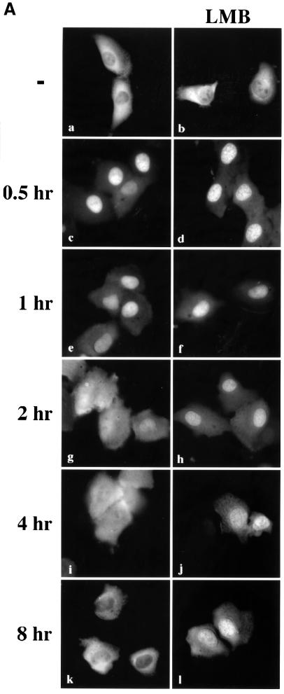
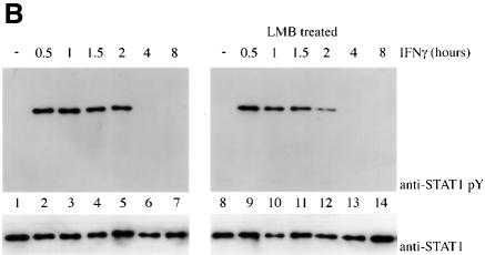
Fig. 2. Leptomycin B inhibits STAT1 nuclear export. (A) U3A cells expressing STAT1–GFP were pre-treated for 30 min with cyclo heximide (all panels) and leptomycin B (LMB) (right panels) and then pulse-treated with IFN-γ for 30 min. The cellular localization of STAT1–GFP was visualized by fluorescent microscopy. Time after IFN-γ treatment is indicated to the left of the panels. (B) Cells from each treatment condition in (A) were collected and immunoprecipitated with anti-STAT1 antibody. Western blots were performed with anti-STAT1 phosphotyrosine antibody (anti-STAT1 pY) (upper panel) or anti-STAT1 antibody (lower panel).

An official website of the United States government
Here's how you know
Official websites use .gov
A
.gov website belongs to an official
government organization in the United States.
Secure .gov websites use HTTPS
A lock (
) or https:// means you've safely
connected to the .gov website. Share sensitive
information only on official, secure websites.

