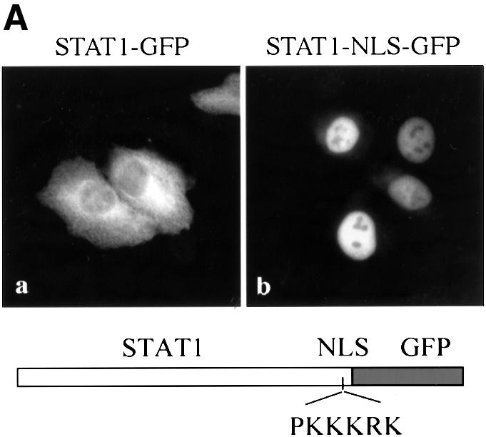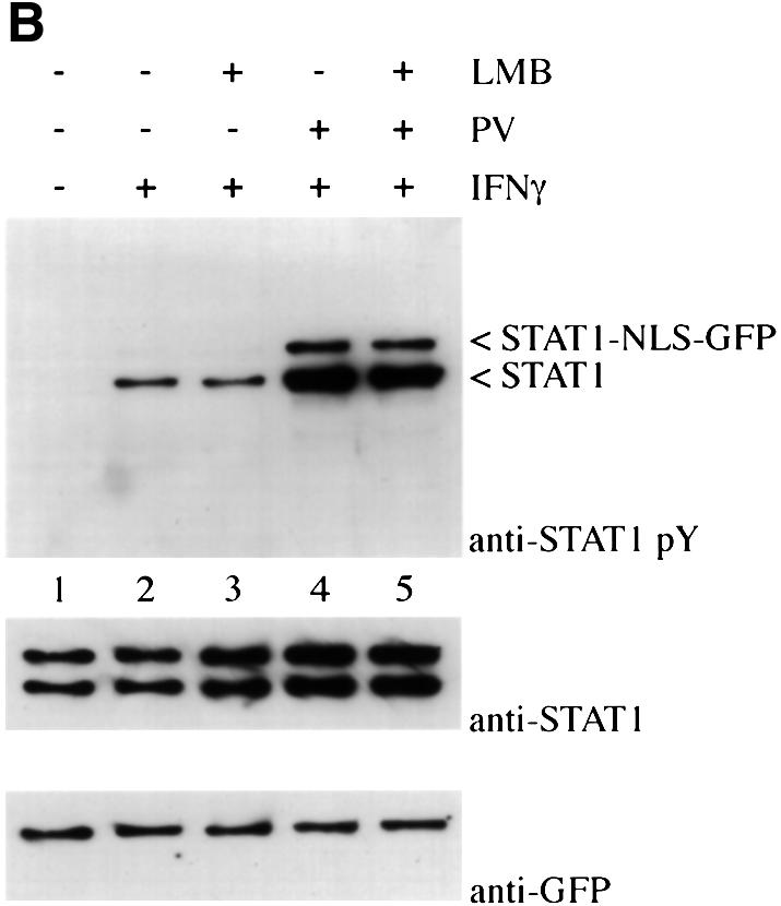

Fig. 8. Effect of addition of a characterized NLS to STAT1. (A) HT1080 cells were transfected with either STAT1–GFP (a) or STAT1–NLS–GFP (b) and localization determined in untreated cells by fluorescent microscopy. (B) HT1080 cells stably expressing STAT1–NLS–GFP were pre-treated with cycloheximide (all lanes) and leptomycin B (LMB) (lanes 3 and 5) for 30 min. Cells were left untreated, or treated with IFN-γ alone or with IFN-γ and pervanadate (PV). Proteins were immunoprecipitated from lysates with anti-STAT1 antibody, and western blots were performed with anti-STAT1 phosphotyrosine antibody (anti-STAT1 pY) (upper panel), anti-STAT1 antibody (middle panel) or anti-GFP antibody (lower panel).
