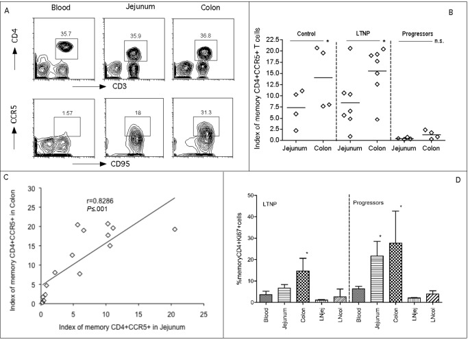
Figure 3.
Comparison of mucosal memory CD4+CCR5+ T cells and proliferation in jejunum and colon. A, memory CD4+CCR5+ T cell expression in blood, jejunum, and colon in a representative normal Chinese rhesus macaque (RM). Cells were first gated on lymphocytes, CD3+ T cells and then CD4+ T cells, and they were then analyzed for CD95+ and CCR5+. B, Comparison of mucosal memory CD4+CCR5+ T cells (Index) between jejunum and colon within the same group. *P < .05. (Data were not available for 1 progressor). P < .05 when compared with the matched sample in the control and long-term nonprogression (LTNP) groups. P < .01 when compared to the matched samples in the LTNP group. The index was previously described elsewhere but reflects the proportion of total CD4+ T cells of the T cell pool (CD3+) that are memory (CD95+) and CCR5+ [21]. C, Correlation of memory CD4+CCR5+ T cells between jejunum and colon in all animals, including groups of control animals, LTNPs, and progressors. D, Proliferation of memory CD4+ T cells in different tissue compartments. *P < .05 compared with peripheral blood specimens within the same group. n.s., no significance.

