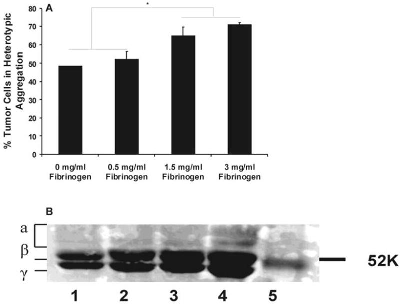
FIGURE 4.

(A) Effect of Fg concentration on PMN-Lu1205 binding. Lu1205 cells were subject to shear with PMNs in the presence of different concentrations of Fg in a cone-plate viscometer at a shear rate of 62.5 s-1. Cell suspensions were fixed with formaldehyde at 60 sec after onset of the experiments. Data represent means ±SEM (n>3). * P<0.05. (B) SDS-PAGE for fibrinogen digested products. Fg was cleaved by thrombin at different levels (0.05 U/ml (Lane 3) and 2 U/ml (Lane 4)) for 10 min. The reaction products were subjected to 12% polyacrylamide gel separation. Separated proteins were stained by Coomassie blue. Lane 1 is fibrinogen, Lane 2 is control and Lane 5 is marker.
