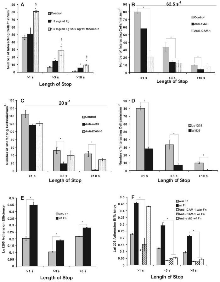
FIGURE 8.

Relative contributions of αvβ3 and ICAM-1 to fibrin(ogen)-mediated PMN-melanoma-EC interaction under flow in parallel plate. (A-D) The number of PMNs contacting with Lu1205 or WM35 cells for more than 1 sec (initial tethering), 3 sec (transient adhesion) and 10 sec (long-term adhesion): (A) Fibrin(ogen) enhanced PMN and melanoma cell interaction. Data are means ± SEM (n>3). *P<0.05 compared with control. § P<0.05 compared with fibrinogen cases; (B-C) Roles of αvβ3 and ICAM-1 in different phases of adhesion of PMNs to Lu1205 cells under shear rate of 62.5 s-1 (B), or 20 s-1 (C). Values are means ± SEM (n>3). *P<0.05 compared with fibrin only cases; (D) PMNs bind weakly to non-metastatic melanoma cell line WM35. Values are means ± SEM (n>3). *P<0.05 compared with Lu1205 cases. (E-F) The adhesion efficiency of Lu1205 cells binding to EI cells for more than 1 sec (initial tethering), 3 sec (transient adhesion) and 5 sec (long-term adhesion) as a result of Lu1205 and PMN collisions: (E) Role of soluble fibrin(ogen) on melanoma adhesion efficiency to EI cells via PMNs at shear rate of 62.5 s-1; (F) Roles of αvβ3 and ICAM-1 in Lu1205 adhesion at the shear rate of 62.5 s-1. *P < 0.05 compared with the “Lu1205+Fn” case. Values are mean ± SEM (n>3).
