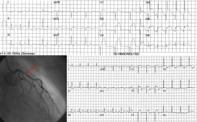
Fig. 1.
Example of a patient. The top ECG shows an acute anterior wall infarction. The patient received active treatment and subsequently underwent angiography which revealed a TIMI III flow with more than 70% occlusion of the proximal LAD. A stent was placed and the ECG 60 min after primary PCI showed more than 70% ST resolution

