FIGURE 4.
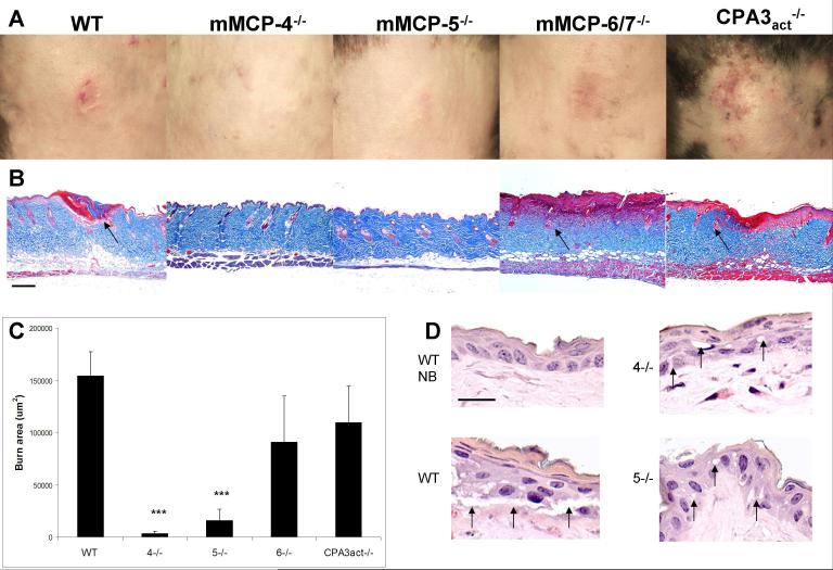
The absence of MC-specific secretory granule proteases, mMCP-4 or mMCP-5, protects against epidermal scald injury. A, The progression of clinical injury to ulceration at d 3 occurs in the WT, mMCP-6/7- and CPA3act-deficient mice, but not in mMCP-4- or mMCP-5-deficient mice. B, The corresponding histological sections after Masson’s trichrome staining to assess the breadth and depth of injury by the appearance of denatured collagen (red), injured hair follicles (arrows), and edema of the dermis revealed by thickness and lighter staining. Each picture is from a single mouse representative of the injury characteristic of each strain. C, Burn injury area (red area of denatured collagen in histological sections) in the same groups as in A. Values for the burn area are the mean (± SE) um2. The value for the WT mice is derived from 3 experiments with 20 mice while the other values are from 1 experiment with 4, 4, 6, and 5 mice, respectively. Statistical values are from a t-test relative to the WT value. D, A high magnification of the epidermis in unburned WT (NB) and in burned WT , mMCP-4−/− (4−/−), and mMCP-5−/− (5−/−) mice at 2 h post burn shows disruption of epithelial tight junctions and epithelial vacuolization (arrows) in the burned WT mice but only minimal changes in these parameters mMCP-4−/− and mMCP-5−/− strains. Additional histologic data showing protection in the mMCP-4- and mMCP-5-deficient strains are shown in Fig. 5 and quantitated in Fig. 6. Scale bar: B=200 um, D= 20 um.

