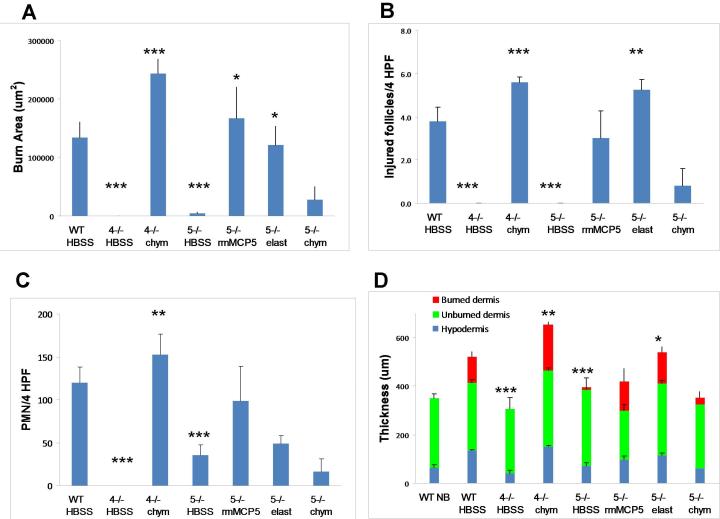
FIGURE 6.
The quantitative assessment of burn injury after the topical application of human chymase to mMCP4−/− mice and of rmMCP-5 or human elastase to mMCP-5−/− mice. Mice were burned for 35 s at 54 °C, ,then treated with topically applied HBSS, chymase (chym), rmMCP-5 or elastase (elast) as indicated for 1 h and the injury evaluated on d 3 post scald burn. A, The mean area (±SE) of burn injury area was assessed by Masson’s trichrome staining. The value for the WT mice is derived from 3 experiments with 16 mice. The value for the HBSS-treated mMCP-5−/− is derived from 2 experiments while all others are from 1 experiment. Numbers of mice are 16, 4, 5, 7, 5, 4, and 5 in each group, respectively. B, The mean number ( ±SE) of injured hair follicles (per 4 HPF) in the same animals as in A. C, The mean number (± SE) of neutrophils (per 4 HPF) identified by CAE reactivity in the hypodermis of the same animals as in A. D, The mean depth (± SE) of the skin from the same mice as in A with the depth of the various individual tissues indicated by burned dermis (red), unburned dermis (green) and hypodermis (blue). The mean (± ½ range) values from 2 non burned WT (WT NB) mice are shown for comparison. For statistical analysis, the mMCP-4- and mMCP-5-deficient mice treated with HBSS were compared to the WT controls, while the enzyme reconstituted groups were compared to the respective HBSS-treated strain. *, p<0.05; **, p<0.01; ***, p<0.001.

