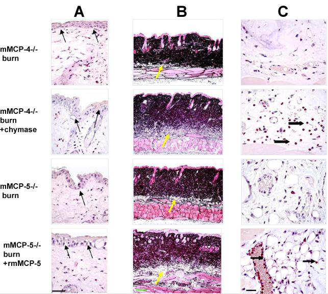
FIGURE 7.
The histological changes 2 h after an epidermal scald in the skin of mMCP-4- and mMCP-5-deficient mice without and with topical application of human MC chymase or rmMCP-5, respectively. All mice were treated with topical HBSS without or without a protease for the first h post burn. A, Cytoplasmic vacuolization and disruption of the tight junctions between the basal cells of the epithelium at the scald site is apparent after CAE in the protease-deficient strains and is accentuated with topical application of chymase to the mMCP-4-deficient and of rmMCP-5 to the mMCP-5-deficient strain (arrows). B, Jones’ staining shows increased edema as indicated by the lighter color of the dermis and greater depth of the hypodermis (yellow arrows) in mMCP-4−/− mice treated with chymase, and of mMCP-5−/− mice treated with rmMCP-5 relative to their HBBS-treated protease-deficient controls. C, The CAE reactivity in the hypodermis shows neutrophils (arrows) in the blood vessels and tissue of mMCP-4−/− mice treated with chymase, and in mMCP-5−/− mice treated with rmMCP-5. Scale bar: A=50 um, B=200 um, C= 25 um.

