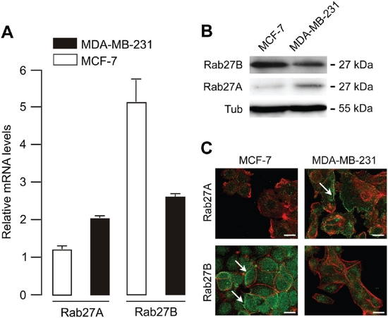
Figure 1: Rab27A and Rab27B mRNA and protein expression in MCF-7 and MDA-MB-231 breast cancer cells.
(A) Rab27A and Rab27B mRNA expression detected via quantitative real-time PCR (relative to human mammary epithelial cell line MCF10A). To demonstrate Rab27A and Rab27B mRNA expression we combined 50 ng cDNA, Taqman gene expression master mix reagent and Assays-On- Demand (Applied Biosystems, Austin, TX) for Rab27B (Assay ID Hs00188156_m1), Rab27A (Assay ID Hs00608302_m1), and the household gene PIAA (Assay ID Hs99999904_m1) on an ABI PRISM 7900 HT Sequence Detection System (Applied Biosystems) using the 2−ΔΔCT method for relative gene expression. The cycling conditions comprised 2 min at 50°C, 10 min at 95°C and 40 cycles at 95°C for 15 s and 60°C for 60 s. (B) Rab27A and Rab27B protein expression detected via Western blot analysis using the specific goat polyclonal Rab27A antibody (C-20, Santa Cruz Biotechnology) and our specific rabbit polyclonal Rab27B antibody. (C) Laser scanning confocal images showing endogenous localization of Rab27A and Rab27B (green) and F-actin (red) in MDA-MB-231 and MCF-7 breast cancer cells. Scale bar: 20 μm.

