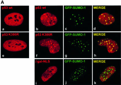
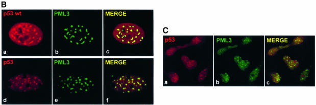
Fig. 1. p53 relocalizes into NBs. (A) SaOS-2 cells were microinjected with either 10 ng/µl pcDNA3p53wt (b–d), pRcCMVp53K386R (f–h) or pNLS-βgal (i–k) together with 30 ng/µl pGFPSUMO-1 and 50 ng/µl pcDNA3HAhUbc9. p53 staining was analysed using a rabbit antiserum, while the localization of βgal-NLS was detected by a monoclonal anti-β-galactosidase antibody. Primary antibodies and GFP–SUMO-1 staining were revealed by incubation with TRITC-conjugated secondary antibodies or by the intrinsic green fluorescence of GFP, respectively. Merging of the two colours results in a yellow signal, corresponding to colocalized proteins. (a and e) Staining of p53 wt and K386R, respectively, in the absence of coexpressed GFP–SUMO-1 and HA-hUbc9. (B) SaOS-2 cells (a–c) microinjected with 10 ng/µl pcDNA3p53wt and 30 ng/µl pcDNA3PML3 were analysed for p53 expression as above and for PML staining using the anti-PML monoclonal antibody PG-M3 followed by incubation with an FITC-conjugated secondary antibody. U2OS cells (d–f) were microinjected with pcDNA3PML3, and localization of endogenous p53 and overexpressed PML3 was examined as above. (C) LOVO cells were treated with UV light and As2O3 and expression of endogenous p53 was detected by a mixture of DO-1 and 1801 monoclonal antibodies followed by incubation with a TRITC-conjugated secondary antibody (a and c). Endogenous PML3 staining was revealed with a rabbit polyclonal serum specific for PML3 and FITC-conjugated secondary antibody (b and c).

An official website of the United States government
Here's how you know
Official websites use .gov
A
.gov website belongs to an official
government organization in the United States.
Secure .gov websites use HTTPS
A lock (
) or https:// means you've safely
connected to the .gov website. Share sensitive
information only on official, secure websites.

