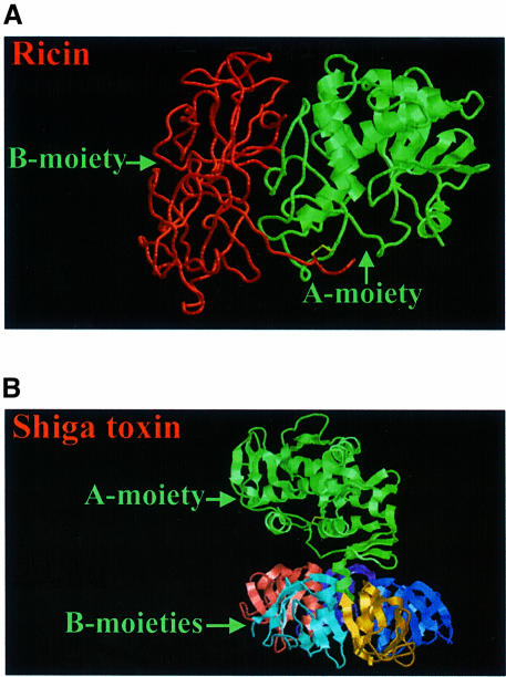
Fig. 2. Crystallographic structures of ricin (A) and Shiga toxin (B). The enzymatically active subunits are in green, whereas the binding moiety of ricin is in red, and the five small binding subunits of Shiga toxin are multicoloured. The disulfide bond (and the neighbouring carbon atoms) connecting the two chains of ricin, and the internal disulfide bond in the A-moiety of Shiga toxin are yellow. The structures have been obtained from the PDB protein data bank (ricin: 1DMO; Shiga toxin:2AA1), and are based on work published by Rutenber et al. (1991) and Fraser et al. (1994).

An official website of the United States government
Here's how you know
Official websites use .gov
A
.gov website belongs to an official
government organization in the United States.
Secure .gov websites use HTTPS
A lock (
) or https:// means you've safely
connected to the .gov website. Share sensitive
information only on official, secure websites.
