
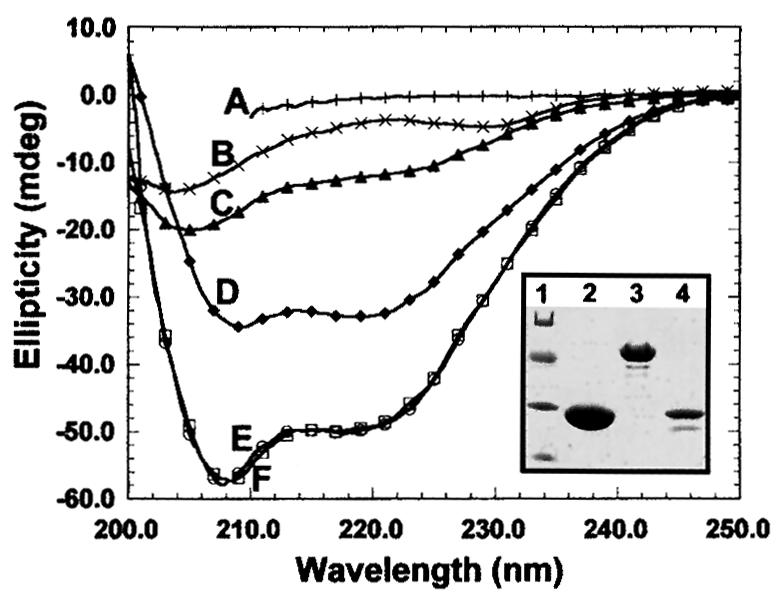
Fig. 2. Structural features of recombinant M31. Circular dichroism spectra of purified His6-M31 (C), GST (D) and M31–GST (E).Curve F is the sum of the His6-M31 and GST spectra. The profile of fully denatured M31–GST is depicted in A. Plot B corresponds to the spectrum of the M31 peptide His6-M31N (for more information on this see Figure 8). The inset shows the electrophoretic mobility of the analyzed proteins in SDS–PAGE gels. Lane 1, molecular weight markers with values of 68, 41, 31 and 21 kDa; lane 2, pure GST; lane 3, M31–GST; lane 4, His6-M31.