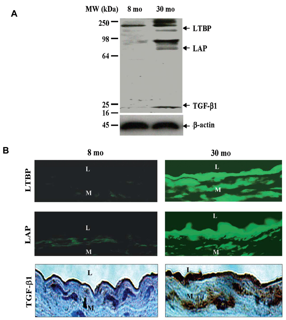
Figure 8. Rat aortic TGF-β1 protein expression.
A. Western Blots for TGF-β1. B. Immunofluorescence staining for LTBP (upper panels, FITC, green color) and LAP (middle panels), and immunohistochemical staining for TGF-β1 (lower panels, DAB, brown color). L=lumen; M=media. From Wang M et al [7].

