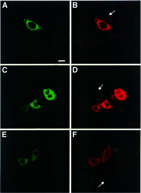
Fig. 2. Intracytoplasmic expression of p34 and p58. Confocal immunofluorescence microscopy of HEp-2 cells transfected with DNA encoding GFP–p34 (A), GFP–p58 (C) or GFP–VacA (E). Expressed proteins were stained with antibodies directed against p34 (B) and the C-terminal part of VacA (D and F). Arrows indicate the presence of non-transfected cells not reacting with specific antibodies. Bar, 10 µm.

An official website of the United States government
Here's how you know
Official websites use .gov
A
.gov website belongs to an official
government organization in the United States.
Secure .gov websites use HTTPS
A lock (
) or https:// means you've safely
connected to the .gov website. Share sensitive
information only on official, secure websites.
