Figure 4.
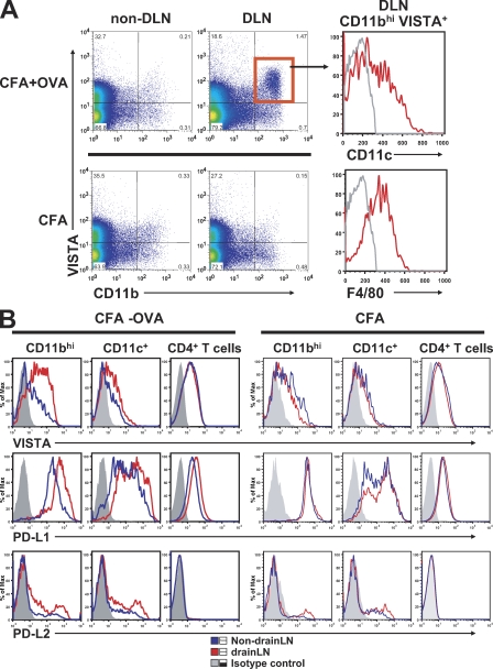
Comparison of in vivo expression patterns of VISTA and B7 family ligands PD-L1 and PD-L2 during immunization. DO11.10 TCR transgenic mice were immunized with 200 µg chicken OVA emulsified in CFA or CFA alone on the flank. Draining LN (DLN) and nondraining LN cells were collected after 24 h and analyzed by flow cytometry for the expression of VISTA, PD-L1, and PD-L2. (A) Representative VISTA expression profile on CD11b+ monocytes at 24 h after immunization. (B) Expression of VISTA, PD-L1, and PD-L2 on CD11bhi monocytes, CD11c+ DCs, and CD4+ T cells was analyzed at 24 h after immunization. Shown are representative results from at least four independent experiments.

