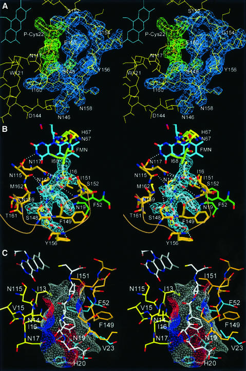
Fig. 6. Substrate binding. (A) 2Fo – Fc electron density contoured at 1σ for the substrate pentapeptide is shown in green, that of the substrate recognition clamp in blue. (B) The substrate peptide (P-Ser19 to P-Cys22) forms a regular parallel β-sheet with β-strand S7 of the substrate clamp (Phe149 to Ile151) and backbone H-bonds to the protein surface on the opposite side to Asn14 (carbonyl and amide of P-Tyr20) and Asn117 (amide and carboxylate of P-Cys22). (C) Surface representation of the binding pocket for P-Tyr20 coloured according to the H-bonding properties of the contributing protein residues (grey, hydrophobic; red, H-bond donors; blue, H-bond acceptors).

An official website of the United States government
Here's how you know
Official websites use .gov
A
.gov website belongs to an official
government organization in the United States.
Secure .gov websites use HTTPS
A lock (
) or https:// means you've safely
connected to the .gov website. Share sensitive
information only on official, secure websites.
