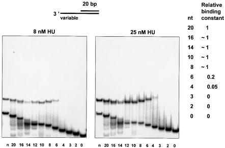
Fig. 4. HU-binding DNA 3′-overhangs of different lengths of the ss-part (indicated at the bottom in nt), or DNA containing a nick (indicated as n), were analyzed. HU protein was added to a final concentration of 8 (left) or 25 nM (right) to 0.05 pmol of labeled DNA as described in Materials and methods. Bound and free DNA were gel-separated, and binding constants of HU–DNA complexes were calculated. The binding constant of the 20-nt 3′-overhang was taken as 1 to calculate relative affinities of overhangs of different sizes, which are indicated on the right.

An official website of the United States government
Here's how you know
Official websites use .gov
A
.gov website belongs to an official
government organization in the United States.
Secure .gov websites use HTTPS
A lock (
) or https:// means you've safely
connected to the .gov website. Share sensitive
information only on official, secure websites.
